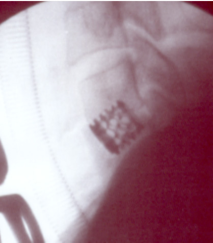
An intraoperative, positive image X-ray of the patient's neck taken from the side with the head at the top, feet at the bottom, front to the left and back to the right.

Salient features that are visible are the ribbed anaesthetic tube on the left traversing the trachea down towards the lungs, the chunky blocks of the vertebral bodies in the centre stacked one upon the next, their spinous processes on the right angling downwards and backwards, and the small titanium cage firmly embedded between the two lower vertebral bodies visible.

In time, it is intended that bone should grow through the cage and its lattice structure, fusing it and the two adjacent vertebral bodies together permanently.
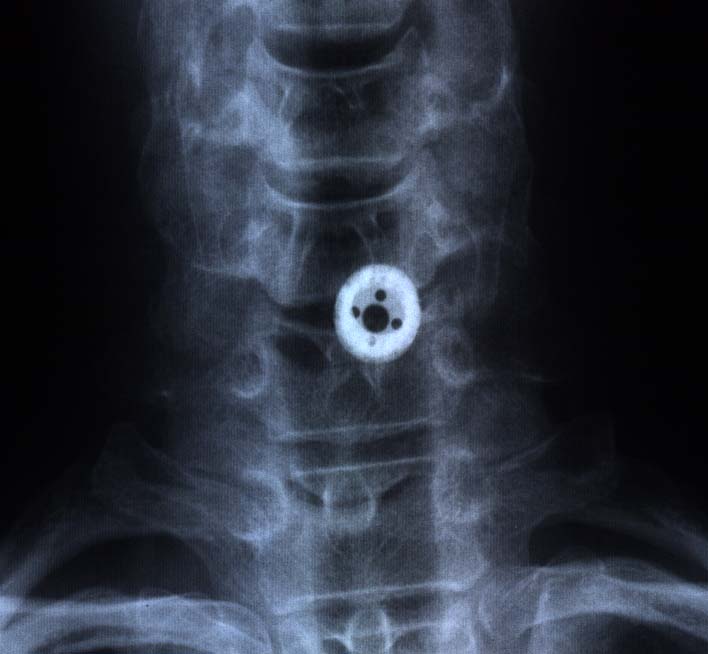
And a month after the operation, a true X-ray taken from the front shows the cage in good position, slightly off-centre, but holding the two vertebral bodies apart. Although not obvious in this view, the bone chips which were placed within the cage have started the process of fusing the two bodies together with a continuous bridge of bone which ultimately will engulf the cage completely and form a solid double-vertebra.


To return to the previous page please close this browser window